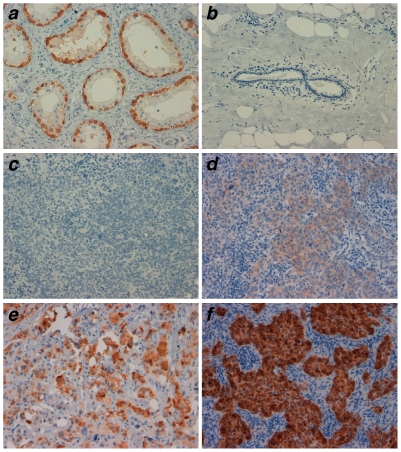
Figure 5. Expression of ESO in triple negative BC.
Expression was assessed by IHC staining of paraffin-embedded tumors using the ESO-specific mAb E978. Tumors were scored based on the percentage of positive cells (-, 0-rare; 1, <10%; 2, 10–25%; 3, 25–50%; 4, >50%) and the staining intensity (A, faint; B, moderate; C, strong). Testis (positive control, a), normal breast (negative control, b) and examples of triple negative tumors scored as negative (c), 4B (d), 3C (e) and 4C (f) are shown. Magnification: ×20.

