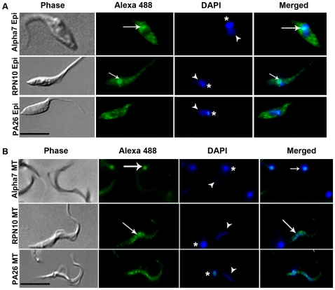
Figure 2. Intracellular localization of proteasomal proteins.
Parasites were labeled with antibodies specific to the catalytic subunit 7 of 20S proteasome (alpha 7) regulatory subunit 10 of 19S (RPN10) and activator proteasome 26 (PA26) in (A) three-day-old cultured epimastigotes (Epi) and (B) metacyclic trypomastigotes (MT). DAPI were used to stain nuclei (arrow head) and kinetoplast (asterisk). Merged images suggesting co-localization of alpha 7 (20S proteasome) with kinetoplast (arrow) of epimastigotes and mainly metacyclic trypomastigotes and co-localization of RPN10 (19S complex) with nuclei (arrow) of epimastigotes and metacyclic trypomastigotes and co-localization of RPN10 (19S complex). Bars = 10 µm.

