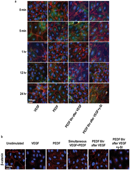
Figure 3. PEDF prevents VEGF-induced dissociation of endothelial AJs and TJs.
Representative immunofluorescent images of confluent cultures of microvascular endothelial cells treated with vehicle (unstimulated), VEGFA alone, PEDF alone, simultaneous VEGFA+PEDF, PEDF 6 hours post VEGF and PEDF 6 hours post VEGF+γ-secretase inhibitor (γ-SI) (n = 4 independent experiments). VEGFA and PEDF were used at 100 ng/ml and γ-secretase inhibitor at 1 nM. (a) Cultures were triple stained for VE-cadherin (red), claudin-5 (green) and nuclei (DAPI, blue) and assessed at different times over 24 hours using confocal microscopy. Merged images are shown with colocalization of VE-cadherin and claudin-5 appearing as yellow. (b) The effect of PEDF on β-catenin using the conditions described in (A) (n = 4). Scale bar = 100 µm.

