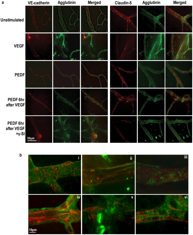
Figure 5. PEDF prevents VEGF-induced dissociation of endothelial AJs and TJs in the retinal vasculature of mice.
(a) Representative confocal images of retinal vessels in flat mount preparations from unstimulated and animals, treated as described in Figure 4, immunostained with VE-cadherin or claudin-5 (red) and FITC-conjugated agglutinin (green) to visualize retinal vessels. Merged images are shown with colocalization of VE-cadherin and claudin-5 appearing as yellow. Scale bar = 50 µM. The lower panel (b) shows representative merged higher power images of retinal vessels stained for VE-cadherin (i–iii) or claudin-5 (iv–vi) (red) and FITC-conjugated agglutinin (green). i, iv = unstimulated; ii, v = VEGF treatment; iii, vi = PEDF 6 hr after VEGF. Scale bar = 10 µM.

