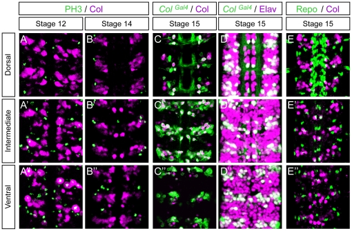
Figure 1. Col is expressed in neurons but not in glial cells in the embryonic Drosophila VNC.
(A–A″) Col expression (magenta) relative to mitotic pattern as detected by Phospo-Histone H3 (PH3, green). At stage 12, two to three PH3/Col expressing cell are typically found per hemisegment in the ventral region of the VNC. (B–B″) At stage 14 expression of Col does not overlap with PH3 indicating that Col is only expressed in postmitotic cells. (C–C″) At stage 15 Col expression overlaps extensively with expression of ColGal4. (D–D″) At stage 15 ColGal4 reveals that Col expressing cells are neurons (Elav+). (E–E″) At stage 15 no overlap between Col and Repo is found showing that Col is not expressed in glial cells. Dorsal, Intermediate and Ventral indicate the different focal planes of the VNC. In all panels three consecutive segments are shown. Anterior is upwards in all panels.

