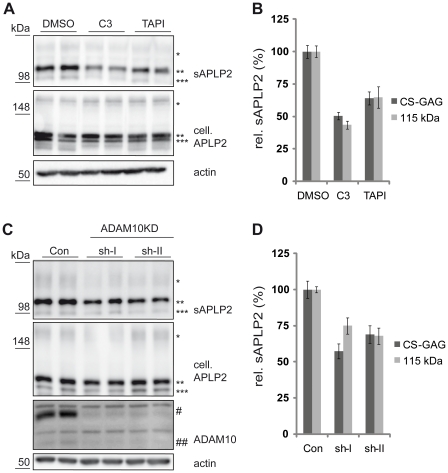
Figure 3. APLP2 processing in primary cortical neurons.
(A) E16 primary cortical neurons were treated with C3 (1 µM), TAPI-1 (50 µM) or DMSO as a solvent control. sAPLP2 (* CS-GAG-modified, ** 115 kDa, *** 100 kDa) was detected in the conditioned medium of these cells. Full-length cellular APLP2 (cell. APLP2) and actin were detected in the cell lysate. A clear reduction of sAPLP2 levels was observable upon C3 as well as TAPI-1 treatment. (B) Quantification of experiments in A (mean +/− SEM). C3 and TAPI-1 significantly reduced sAPLP2 (C3: p<0.001 for both detected species, TAPI-1: p<0.001 for CS-GAG modified APLP2, p = 0.004 for mature APLP2; n = 6). (C) E16 primary cortical neurons with lentiviral knock-down of ADAM10 were analyzed. shRNAs (sh-I and sh-II) targeting different regions of ADAM10 and a non-targeting shRNA control (Con) were used. A clear reduction of ADAM10 (# immature, ## mature) levels was observed in the cell lysate and of sAPLP2 in the conditioned medium. (D) Quantification of experiments in C (mean +/− SEM). Both shRNAs significantly reduced sAPLP2 (p<0.01 for both detected species for both shRNAs, n = 6).

