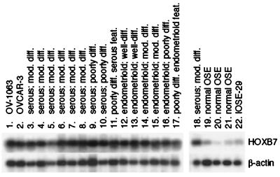
Figure 3.
Semiquantitative RT-PCR analysis of HOXB7 expression. Shown are Southern blots of HOXB7 and β-actin RT-PCR products in OV-1063, OVCAR-3 and IOSE-29 cells (lanes 1, 2, and 22), specimens of normal OSE (lanes 19–21) and ovarian carcinomas (lanes 3–18). Histology of carcinomas ranged from poorly differentiated (diff.) with either serous or endometrioid features (feat.) to moderately (mod.) and well-differentiated serous and endometrioid. The specimen used for analysis shown in lane 18 is the same as that in lane 6.

