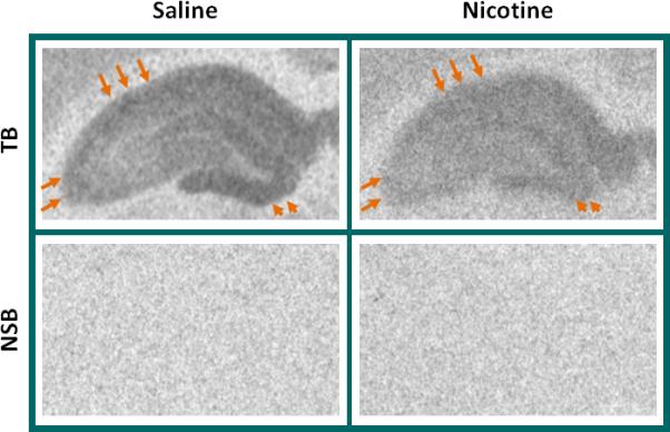
Figure 7. Prenatal nicotine exposure continues to regulate [3H]AMPA binding to AMPAR in the young adult.

Rats were treated with saline (control) or 4mg/kg/day nicotine in utero and on P63, tissues were processed for autoradiography of [3H]AMPA binding, used to detect the distribution of functional AMPAR. A lower density of [3H]AMPA binding is evident throughout the hippocampus of nicotine-exposed pups (right) compared to those exposed to saline (left). Arrows point to CA1 (top) and CA3 (left) areas that show particularly high binding of [3H]AMPA in controls (left) but that were depressed after prenatal nicotine (right). Arrowheads indicate the dentate gyrus. Nonspecific binding (NSB) was assayed in adjacent sections and is shown as background directly below the total binding (TB) image for that animal. All images are from the same film.
