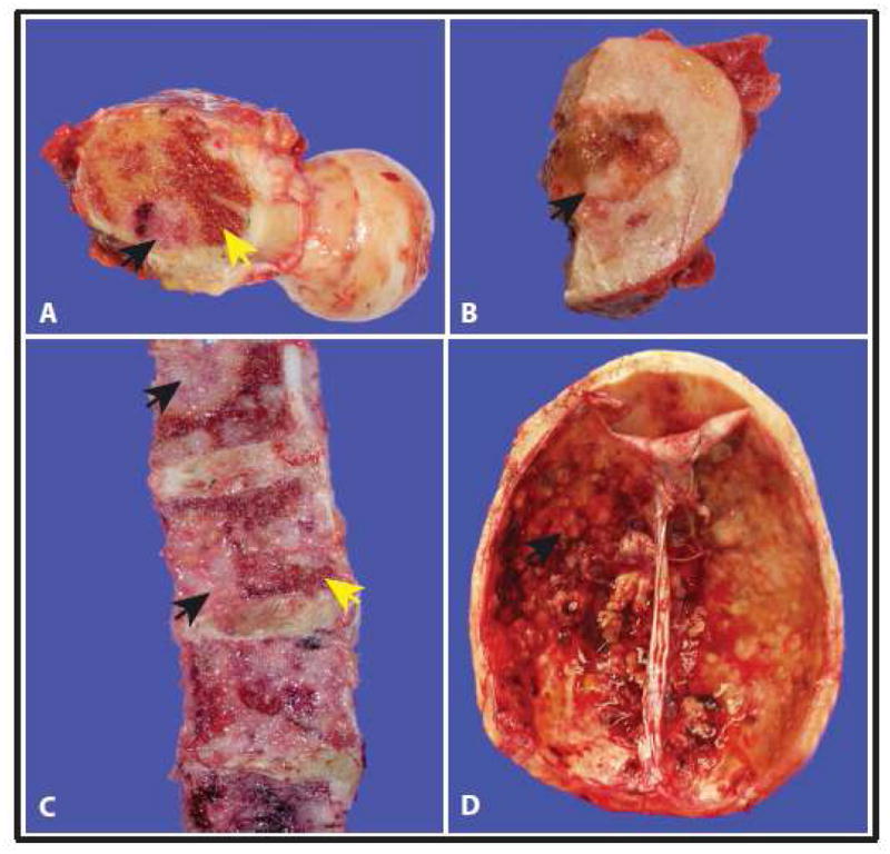
Figure 2.

Gross representation of bone metastasis at different sites from our warm autopsy series of androgen independent prostate cancer patients. (A) and (B) Femoral head (C) Vertebrae (D) Skull and dura. For each image, black arrows indicate metastatic tumor foci and yellow arrows indicate residual bone marrow.
