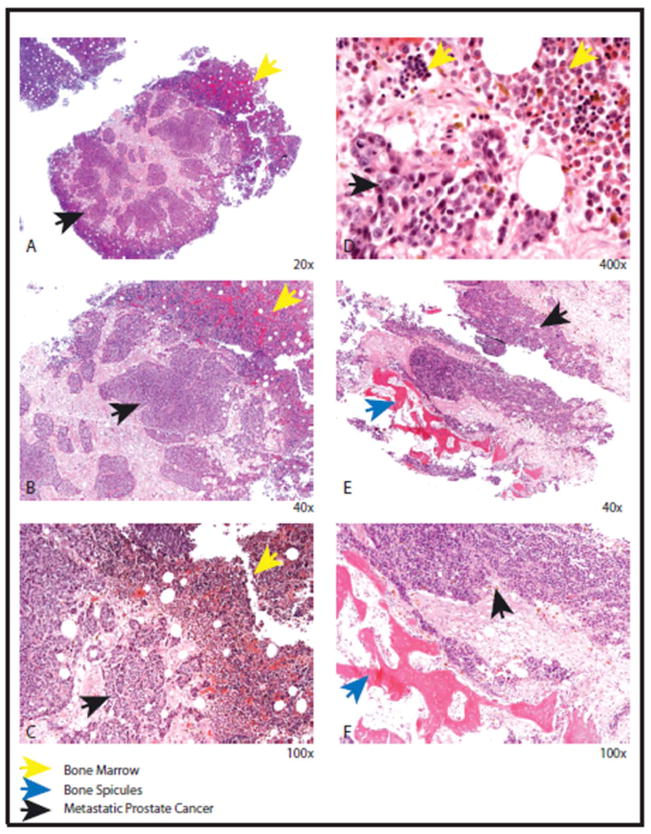
Figure 3.
Histopathology of androgen independent metastatic osseous metastases. Solid sheets and nests of uniform tumor cells indicated by black arrow (A, B, C and D) adjacent to residual normal bone (yellow arrow). The tumor cells have round nuclei with prominent nucleoli compared to the residual bone with normal hematopoietic maturation (D). The tumor cells (black arrow) can be seen adjacent to the residual bone spicules (blue arrow) ( E and F).

