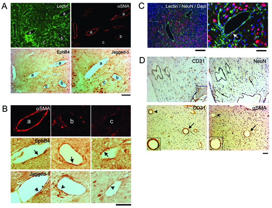
Figure 2. Dysplastic vessels have irregular smooth muscle coverage and express inconsistent arterial and venous markers.
(A) A group of dilated irregular vessels labeled with lectin (green). Some of these vessels have smooth muscle coverage (αSMA, red), some do not. Arterial (Jagged-1) and venous (EphB4) markers were expressed by the endothelial cells of vessels with or without smooth muscle. Scale Bar: 50 µm.
(B) Enlarged image of the vessel (a), (b), (c) indicated in (A). Vessel (a) is covered with a complete layer of smooth muscle and (b) has a few αSMA positive cells. Their endothelial cells express EphB4 and Jagged-1 (arrows and arrowheads). Vessel (c) has almost no αSMA positive cells. Most of its endothelial cells express EphB4, a few express Jagged-1. Scale Bar: 50 µm.
(C) Neuronal tissue is present between enlarged irregular vessels. Arrow indicates NeuN positive cell on the dysplastic vessel wall. Scale Bars: 200 µm (left) and 50 µm (right). Corresponding colors for Lectin, NeuN, and Dapi are green, red, and blue, respectively.
(D) Paraffin sections of surgical specimen from patient shown in Figure 1, stained with antibodies specific to CD31, αSMA or NeuN. Top panel shows NeuN positive cells (arrows) detected between the dysplastic vessels. Lower panel shows enlarged vessels with (arrows) or without (arrowheads) smooth muscle layers. Inserts in the lower panel show enlarged images of the vessels indicated by arrows with endothelial cells stained by CD31 antibody and a thin layer of smooth muscle (αSMA) surrounding the endothelial layer. Scale Bar: 50 µm.

