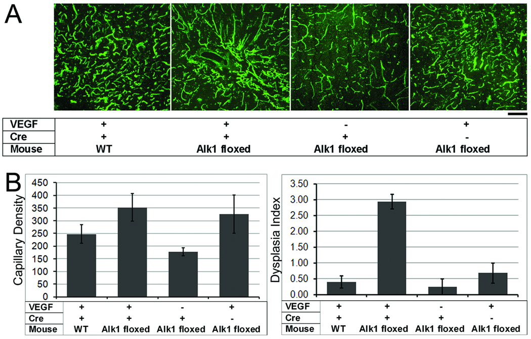
Figure 3. VEGF induced focal angiogenesis in the normal brain and dysplastic vessel formation in the Alk1-deleted brain.
(A) Representative images show lectin-perfused vessels in the brain. Capillary density is high in all AAV-VEGF-treated groups as compared to the AAV-LacZ control group. Dilated irregular vessels were only observed in the brain with Alk1 deletion and VEGF stimulation. Scale Bar=50 µm.
(B) Bar graphs show capillary density (left) and dysplasia index (right). Capillary density increased in all groups with VEGF stimulation as compared to the AAV-lacZ control group (p=0.07). Dysplasia index increased significantly in Alk1-floxed mice treated with Ad-Cre and AAV-VEGF as compared to all other groups (p<0.01).

