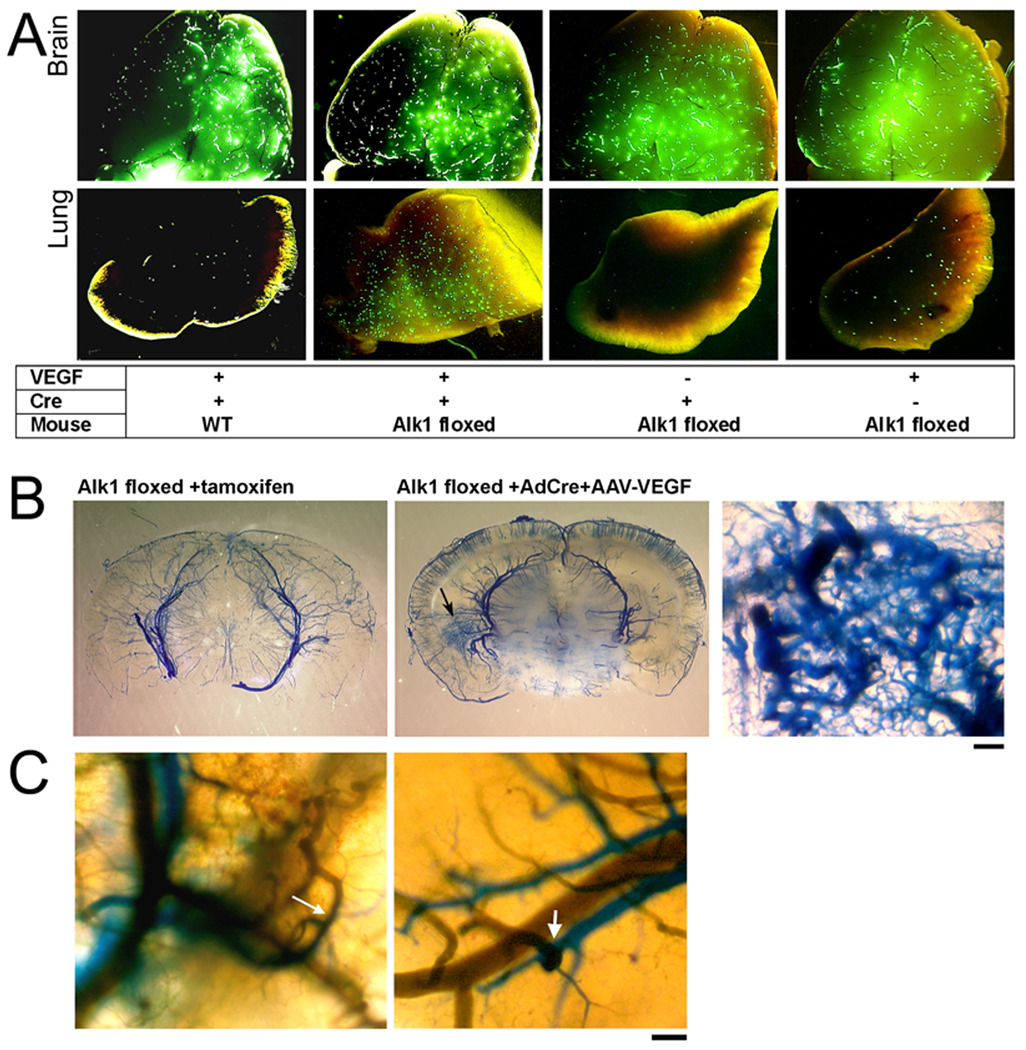
Figure 4. Arteriovenous (A–V) shunting was detected in the brain with regional Alk1 deletion and VEGF stimulation.
(A) Brain and lung samples collected from animals perfused with 20 µm fluorescent beads through the carotid artery. Beads were detected in all brain samples, but only Alk1-floxed mice injected with Ad-Cre and AAV-VEGF had a significant amount of beads in the lung. A few beads were detected in the lungs of control mice injected with AAV-VEGF.
(B) Latex-perfused brain. Latex (blue) presented only in cerebral arteries of Alk12f/2f/ROSA26(+/creER) mice treated with tamoxifen that had Alk1 gene deleted globally (left). Alk1-floxed mice that received intracerebral injection of Ad-Cre and AAV-VEGF had an increase of vascular density at the injection site (arrow, middle image). At higher magnification (right), vessels are enlarged and tortuous with a fistula between arteries and veins. Scale Bar = 100 µm.
(C) Two color latex perfusion shows direct connections of cerebral arteries (blue) and veins (green) (arrows). Scale Bar = 100 µm.

