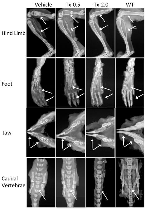
Fig 3.
Representative radiographs of hind limb, foot, jaw bones and caudal vertebrae specimens from 22-day-old Akp2−/− mice treated with vehicle, Tx-0.5, Tx-2.0, and untreated WT mice (radiographic magnification 5X). Arrows indicate improvement in the mineralization of tibia, femur, metatarsals, finger bones, incisors and molars, fracture healing in the tibia and femur, and reduced spaces between adjacent vertebrae of spine in the treated mice compared to the untreated mice.

