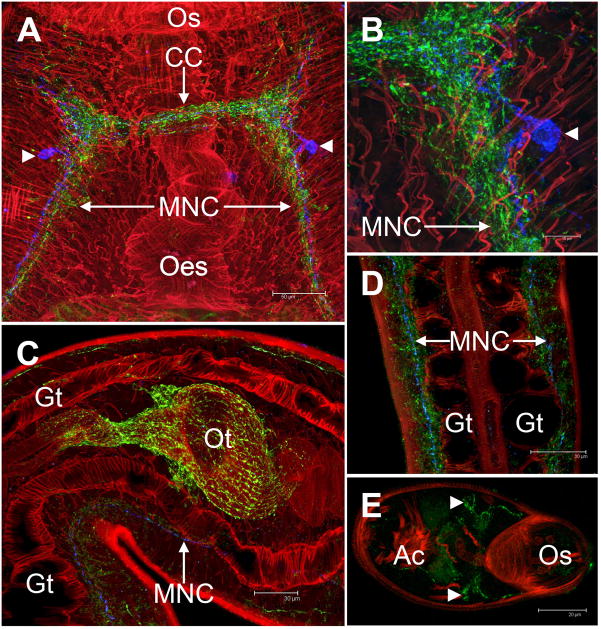
Fig. 2.
Immunocytochemical localisation (ICC) of the Sm-npp-1 neuropeptide GFVRIamide in adult Schistosoma mansoni. (A-D) Blue staining (pseudocoloured Cy5-labelled secondary antibody) indicates GFVRIamide distribution, green (FITC-labelled secondary antibody) represents distribution of FMRFamide-like peptides (FLPs), while red (TRITC; tetrarhodamine isothiocyanate-labelled phalloidin) indicates distribution of filamentous actin; (E) GFVRIamide is denoted by green FITC label. (A) Within the brain of adult S. mansoni, GFVRIamide is expressed in a pair of neurones (arrowheads). GFVRIamide immunoreactivity (IR) projects across the cerebral commissure (CC) and posteriorly along main nerve cords (MNC). (B) One of the bipolar brain neurones expressing GFVRIamide (arrowhead). While GFVRIamide- and FLP-immunopositive nerves occur in close apposition, they never overlap, indicating that they are expressed in separate cells/fibres. (C) FLP-IR is strong in nerves innervating the female ootype (Ot) whereas GFVRIamide-IR appears only in the MNC. (D) Both GFVRIamide- and FLP-IRs are apparent along the entire MNC, running adjacent to the gut (Gt). GFVRIamide-IR is restricted to a single longitudinal nerve tract, while FLP-IR appears in multiple longitudinal nerve tracts in addition to its association with peripheral innervation of somatic muscle layers. (E) In schistosomules, GFVRIamide (stained green) shows a similar expression pattern to adults, localising to neurones associated with each cerebral ganglion (arrowheads) and the longitudinal nerve cords. Ac, acetabulum; OS, oral sucker; Oes, oesophagus.

