Figure 3.
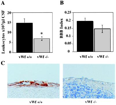
Cytokine-induced meningitis in vWf −/− mice. vWf +/+ (black bars) and vWf −/− (gray bars) mice were inoculated with cytokines via lumbar puncture and with FITC-BSA intravenously 15 min later. Four hours later, cerebrospinal fluid was collected (n ≥ 10). (A) Total leukocyte counts in the CSF were determined. VWf −/− mice had significantly fewer CSF leukocytes. (*, P = 0.005.) (B) Fluorescence in the CSF and the plasma was measured and used to calculate a BBB permeability index. No significant differences were found between vWf +/+ and vWf −/−. (P = 0.12.) (C) Brain tissue was collected and fixed 4 h after meningitis induction. Sections were stained with an antibody to P-selectin. Very strong positive staining was seen on the endothelium of vWf +/+ brains. Very faint staining was present in vWf −/− brain endothelium.

