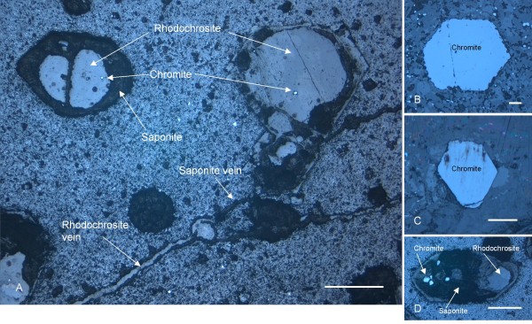
Figure 2.
Optical microphotograph obtained by reflective light. (A) shows the phenocrysts and the mineral succession chromite-rhodochrosite-saponite therein. Veins of rhodochrosite and saponite connecting the phenocrysts are also visible. (B) and (C) show unaltered chromite phenocrysts and their hexagonal and pentagonal shape. (D) shows altered phenocryst with several chromite residues within. Scale bar: (A) 500 μm, (B) 100 μm, (C) 500 μm, and (D) 500 μm.

