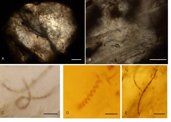
Figure 5.
Optical microphotographs showing the filamentous structures in the rhodochrosite. (A) shows the abundance of filamentous structures in a rhodochrosite filled phenocryst. (B) Close up of filamentous structures showing their smooth, curvi-linear appearance. (C) microphotograph showing a filamentous structure coiled more than 360°. (D) microphotograph showing the twisted, spiral appearance. (E) microphotograph showing branching. Scale bar: (A) 100 μm, (B) 50 μm, (C) 5 μm, (D) 10 μm, (E) 10 μm.

