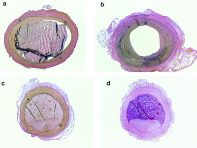
Figure 4.
Examples of carotid arteries of Watanabe rabbits. (a and b) Verhoeft-Van Gieson stain of sections from Watanabe rabbit carotid arteries treated with Ad-TFPI (a) and Ad-RR (b). (c) Plaque in the noninjured contralateral carotid artery of an Ad-TFPI-treated rabbit. (d) Section adjacent to that shown in c and stained with hematoxylin-eosin to illustrate the difference in texture between arterial tissue and latex filled arterial lumen. I, intima; L, lumen filled with residual latex to prevent postmortem distortion of lumen and vessel wall; P, atherosclerotic plaque. →, internal elastic membrane. (×40.)

