Figure 2.
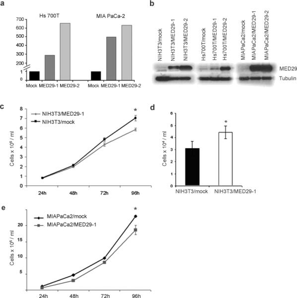
MED29 expression leads to reduced growth of NIH/3T3 cells in vitro. NIH/3T3 mouse fibroblast cell line and Hs 700T and MIA PaCa-2 human pancreatic cancer cell lines were stably transfected with MED29 or an empty control vector. (a) Relative MED29 mRNA expression levels were assessed by qRT-PCR in MED29 transduced Hs 700T and MIA PaCa-2 cells and their respective empty vector (mock) control cells. MED29 expression values were normalized against a TATA-box binding protein house-keeping gene. (b) Western blot was used to detect MED29 protein (21 kDa) in stable MED29-expressing cells vs. mock control cells. β-Tubulin was used as a loading control. (c) The growth of the NIH3T3/MED29-1 and NIH3T3/mock cells was monitored at indicated time points in normal growth medium and (d) after 24h of serum starvation (1% FBS). (e) The growth of the MIAPaCa2/MED29 cells were monitored at indicated time points after release from G1-arrest. The mean +/− s.d. of six replicates are shown. The experiments were repeated three times with similar results. *P < 0.05, **P < 0.005.

