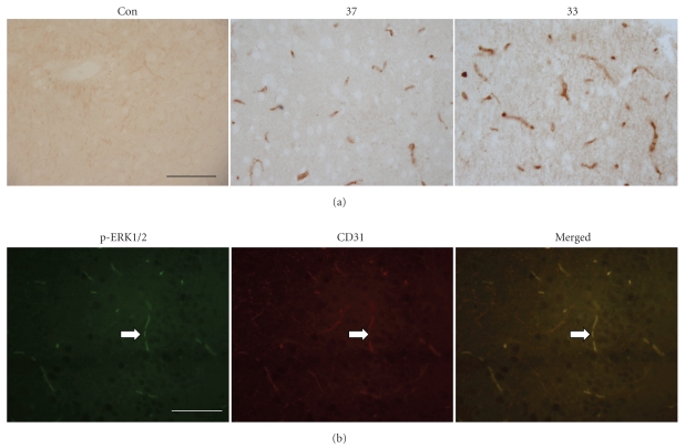
Figure 1.
Photomicrographs of the cerebral cortex in the ischemic brain with immunohistochemical staining for phosphorylated ERK1/2. (a) Phosphorylated ERK1/2 is detected in the ischemic brain under normothermic (37) or hypothermic (33) condition but not in the nonischemic control brain (sham) at 2 hours after MCAO initiation. The number and intensity of ERK1/2 immunoreactive vessels were higher in hypothermic group. (b) Fluorescence double labeling illustrates the colocalization of CD31 (red), an endothelial marker, and phosphorylated ERK1/2 (green) in the vessels of the hypothermia group at 2 hours after MCAO initiation. Scale bar: 100 μm.

