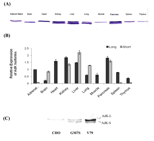
Figure 2.
Differences in expression of two AdK isoforms in various tissues and cell lines. (A) Western blot showing the expression profile of the two isoforms in various rat tissues; (B) Quantification of the relative amounts of two AdK isoforms in rat tissues. Expression levels of the two isoforms were normalized relative to AdK-L level in adrenal gland and the average amount (intensity) ± SD for three independent experiments is shown. (C) Western blot showing relative expression of the two isoforms in CH CHO, V79 and GM7S cell lines.

