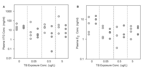
Figure 4.
Comparison of model predictions with measured data in female FHMs exposed to TB for 48 hours. n = 32. White circles represent model predictions, and grey circles represent measured data [39]. Each circle represents one measurement in one fish. (A) plasma VTG concentrations, and (B) plasma E2 concentrations. The x-axis represents TB concentrations in μg/L. Note: for panel B, at 0.5 μg TB/L, there are only 3 measured data points.

