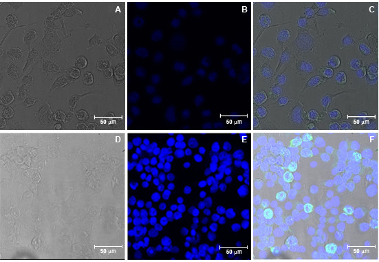
Figure 4.

E protein detection of vSynYFE-infected insect cells by confocal microscopy. Tn5B cells were infected with vSynYFE (10 pfu/cell) and at 72 h p.i., cells were fixed with ice cold acetone and incubated with an E specific monoclonal antibody. Bound monoclonal anti-E antibodies were detected using Alexa488-conjugated anti-mouse antibodies by concofcal microscopy (green). Cell nuclei were visualized by DAPI staining (blue). (A) Tn5B cells mock infected (bright field). (B) Tn5B cells mock infected (DAPI staining). (C) Tn5B cells mock infected (merge). (D) Tn5B cells infected with vSynYFE (brightfield). (E) Tn5B cells infected with vSynYFE (DAPI). (F) Tn5B cells infected with vSynYFE (merge). In A, B, C, D, E and F, Bars = 50 μm. Blue staining (DAPI) indicates cells nuclei.
