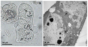
Figure 5.

E protein structural and ultrastructural analysis of vSynYFE-infected insect cells. Tn5B cells were infected with vSynYFE (10 pfu/cell) and at 72 h p.i., the cells were photographed in a light microscope and processed for electon microscopy. Multinucleated syncytial Tn5B cells were easily observed by light (A) and electron microscopy (B). Bar in (A) = 14 μm and in (B), 5 μm. Arrows indicates cells nuclei.
