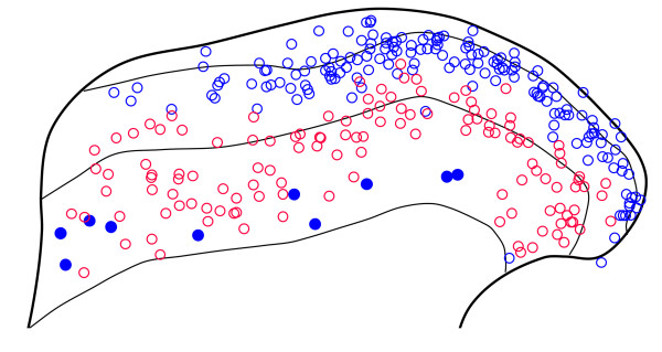
Figure 2.
The distribution of cells that were galanin or parvalbumin immunoreactive in sections reacted to reveal galanin, parvalbumin and nNOS. The diagram shows the laminar location of all of the cells that were galanin (blue) or parvalbumin (red) immunoreactive in the 6 sections that were analysed (2 each from 3 rats). For the galanin cells, those that were also nNOS immunoreactive are shown as filled circles, while those that lacked nNOS are open circles. None of the parvalbumin cells contained nNOS, and there was no colocalisation of galanin and parvalbumin.

