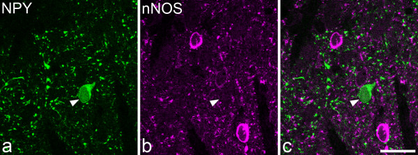
Figure 6.
Lack of co-localisation of NPY and nNOS. A single confocal optical section through lamina II, scanned to reveal: a NPY (green) and b nNOS (magenta). A merged image is shown in c. This field contains cell bodies that are immunoreactive for NPY (arrowhead) or nNOS, but the two types of immunoreactivity are not co-localised. Scale bar = 20 μm.

