Figure 1.
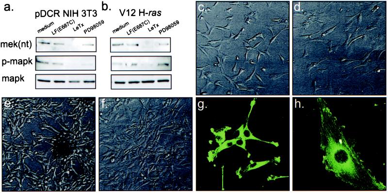
The effects of LeTx on MAPK activation and cell morphology. Immunoblotting of lysates from nontransformed (pDCR NIH 3T3) (a) and V12 H-ras-transformed NIH 3T3 cells (b) show loss of NH2-terminal epitopes of MEK and phospho-epitopes of MAPK after treatment of cells with LeTx but not in cells treated with either medium alone, 100 ng/ml PA plus inactive 10 ng/ml LF(E687C), PA plus LF (100 ng/ml PA plus 10 ng/ml LF), or PD98059 (50 μM from a 50 mM stock in DMSO). Nontransformed cells (c) possessed an irregular, flattened morphology which was not substantially altered by 24 h exposure to LeTx (d). In contrast, after 24 h LeTx treatment, the well defined, elongated, spindle-like shape of V12 H-ras-transformed NIH 3T3 cells (e), reverted to a shape resembling a nontransformed cell (f). Immunostaining of V12 H-ras-transformed NIH 3T3 cells incubated in the absence (g) or presence (h) of LeTx for 24 h for actin (green) showed actin stress fibers formed after LeTx treatment.

