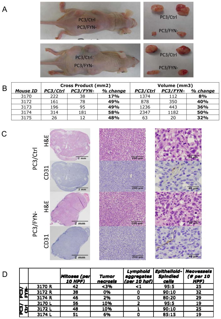
Figure 4.
A) (left panels)Nude mouse with subcutaneous injection of PC3/Ctrl and PC3/FYN- cells at day 57. (right panels) Tumor recovered at necropsy. B) Table of mouse tumor measurements showing both cross products (length x width) and volumetric approximations based on maximal and minimal tumor dimensions. C) Immuohistochemical analysis of tumors from in vivo growth study. PC3/Ctrl (top) and PC3/FYN- cells (bottom) were injected into the flanks of nu/nu mice and collected at necropsy. H&E and CD31 staining is shown at 1× (left panels), 4.2× (middle panels), and 20× (right panels). No significant change in cellular morphology or neovessel formation was seen between the two conditions. D) table of quantification of mitoses, tumor necrosis, lymphoid aggregate formation, ratio of epithliod to spindled cells, and neovessel density. No significant differences were seen.

