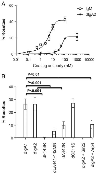
Figure 7.
Binding of IgM and dIgA to hFcα/μR-transfected SGHPL-4 cells assessed by rosette formation. (A) IgM-opsonized erythrocytes bind hFcαμR with greater affinity than erythrocytes opsonized with equimolar concentrations of dIgA2. (B) Rosette formation was assessed with erythrocytes opsonized with dIgA or various dIgA1 point mutants (all at ~333 μg/mL) purified by size exclusion chromatography. Rosette formation by dIgA2-opsonized erythrocytes could be completely ablated by Sir22 and partially by Arp4 when the bacterial IgA-binding proteins were pre-incubated with dIgA2-opsonized erythrocytes at 100 μg/mL. Figures show the mean±SD of two independent experiments. Differences between %rosettes obtained with dIgA1 and the various test groups were analyzed using Dunnett’s multiple comparison test. Statistically significant differences between the groups are indicated.

