
Figure 8.
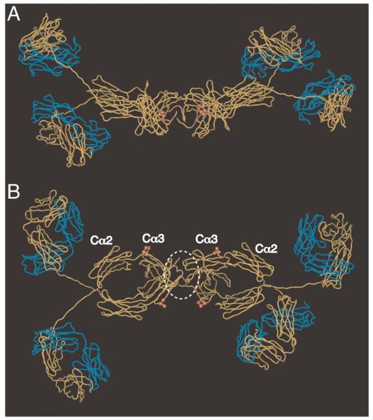
Molecular model of dIgA1 showing residues predicted to be critical for interaction with hFcα/μR. The figure shows two views of the solution structure of human dIgA1 [24] (PDB accession: 2qtj) in which the heavy chains are shown in yellow, and the L chains in blue. The tailpieces are omitted for clarity. The heavy chain residues Pro440–Phe443 implicated in hFcα/μR interaction are shown as red spheres. J chain is not included in the model, but we have added a white dotted line to indicate a possible position. (A) side view; (B) view from above.