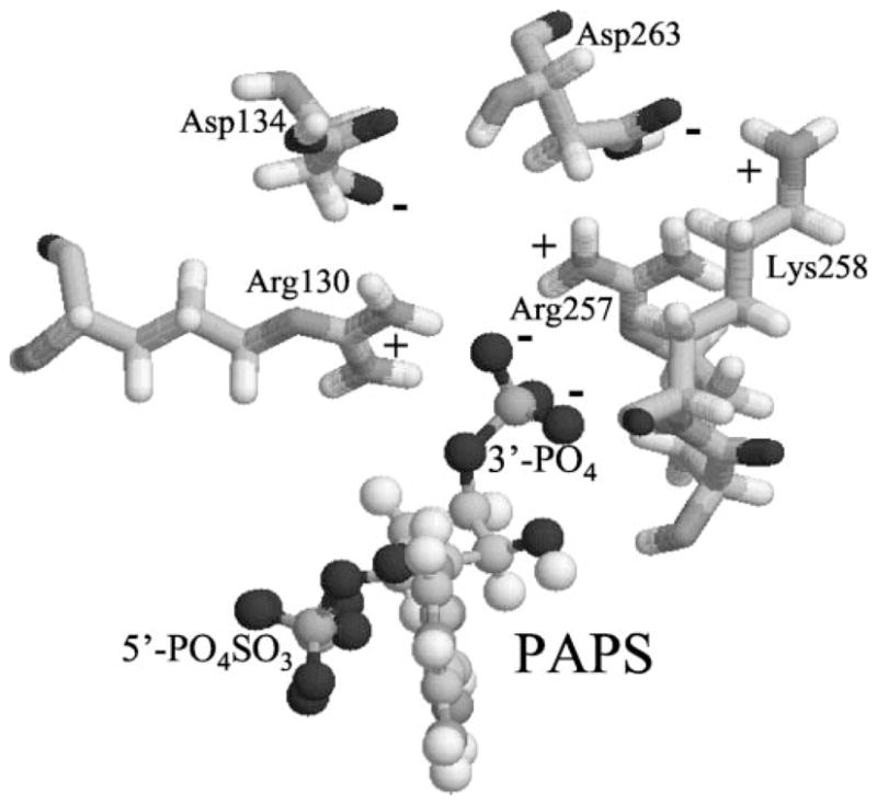
Fig. 9. Computer modeling structure of SULT1A1 showing charged residues in close proximity to 3′-phosphate of PAPS.

The model structure of SULT1A1 was depicted by RasMol 2.6. Only PAPS and charged residues within 5 Å to 3′-phosphate are displaced.

The model structure of SULT1A1 was depicted by RasMol 2.6. Only PAPS and charged residues within 5 Å to 3′-phosphate are displaced.