Figure 5.
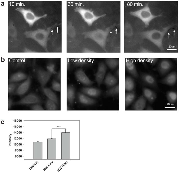
Selective stimulation by TNF α-coated nanowires. a, HeLa cells expressing p65-GFP at the indicated times following stimulation with TNF α-coated nanowires. The lower cell contacts two nanowires (arrows) and exhibits p65 nuclear translocation whereas the upper cell contacts no nanowires and exhibits no translocation. (See also Supplementary Movie 5.) b,c, Wildtype HeLa cells stained for NF-κB following exposure to solutions of 0 (control), 0.2 × 10⁶ (low density), or 1 × 10⁶ (high density) TNF α-coated nanowires/mL, corresponding to 0, ~0.3, ~1.5 nanowires/cell on average, respectively. In (b), representative images are shown. In (c), the average NF-κB level is plotted (more than 150 cells measured for each condition). NF-κB levels were significantly different (p < .001) between the high and low stimulation cases.

