
Fig. 1.
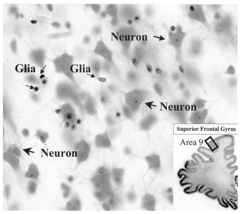
High power micrograph of neurons and glial cells as they appear in the cortical layers of area 9 in coronal section through the prefrontal cortex stained with cresyl violet. Note that both the nucleus and cytoplasm are visible in neurons, while only the nucleus is visible in glial cells. The inset at the bottom right is a low power image of the cresyl violet-stained section containing the spot (black rectangle in area 9) where the high power micrograph was taken. High power: 40× objective; low power: 2× objective.