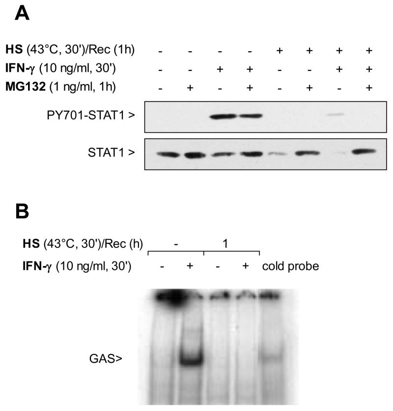
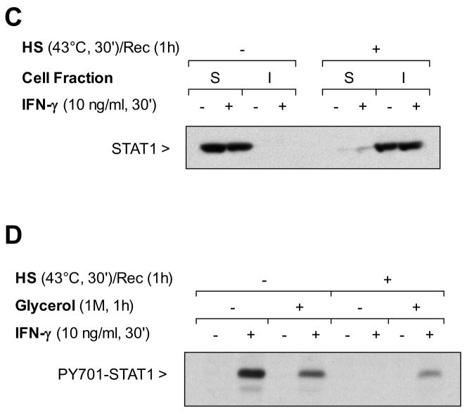
Figure 2. SPR activation prevents IFN-γ activation of the STAT1 signaling pathway, promoting STAT1 detergent insolubility and degradation. Both time and glycerol restore STAT1 signaling after SPR activation.
Panel A. SPR activation prevents phosphorylation of STAT1 in response to IFN-γby causing STAT1 degradation via the proteasome. MH-S cells were either untreated or were incubated at 43°C and allowed to recover for one hour prior to IFN-γ stimulation (10 ng/ml for 30 min) and then analyzed for STAT1 and phosphorylated STAT1 by western blot. Some cells were pretreated with a proteasome inhibitor MG132 (10 μM) one hour prior to exposure to IFN-γ. Panel B. SPR activation prevents the translocation of IFN-γmediated STAT1 to the nucleus. MH-S cells were either untreated or were incubated at 43°C and allowed to recover for 1 hour prior to IFN-γ stimulation (10 ng/ml for 30 min). Electrophoretic mobility shift assay (EMSA) analysis was used to examine STAT1 activity in IFN-γ-treated cells. Nuclear extracts were prepared (see methods) and 5 μg of nuclear protein were incubated with a 32P-labeled DNA containing STAT1 binding consensus sequence (GAS). In some reactions, a 100-fold excess of the unlabeled oligonucleotide was added (indicated as cold probe in the figure). The experiment was performed 5 times. One representative experiment is shown. Panel C. SPR activation causes STAT1 partition into the detergent insoluble cell fraction. MH-S cells were either untreated or were incubated at 43°C and allowed to recover for one hour. Some cells were the treated by IFN-γ (10 ng/ml) or its vehicle for 30 min. Afterwards, the cells were harvested in phosphate buffered saline supplemented with 0.1% Triton X-100, 5 mM MgCl2 and protease inhibitors. The lysates were clarified by centrifugation at 16,000 x g for 10 minutes at 4 C. The supernatant was removed and adjusted to 1x Laemmli sample buffer and the material present within the detergent insoluble pellet was similarly resuspended in 1x Laemmli sample buffer. Aliquots of the detergent insoluble material (I) and the detergent soluble material (S) were analyzed for the presence of STAT1 antibody by Western blot. In each case, the same amount of total protein was applied to the gel. Panel D. Pretreatment with glycerol blocks the heat stress-dependent inhibition of STAT1 phosphorylation in response to IFN-γ . MH-S cells were left untreated or treated with glycerol (1M) for one hour. MH-S cells were either untreated or were incubated at 43°C and allowed to recover for one hour. Cells were the treated by IFN-γ (10 ng/ml) or its vehicle for 30 min and then analyzed for phospho-STAT1 by Western blot.


