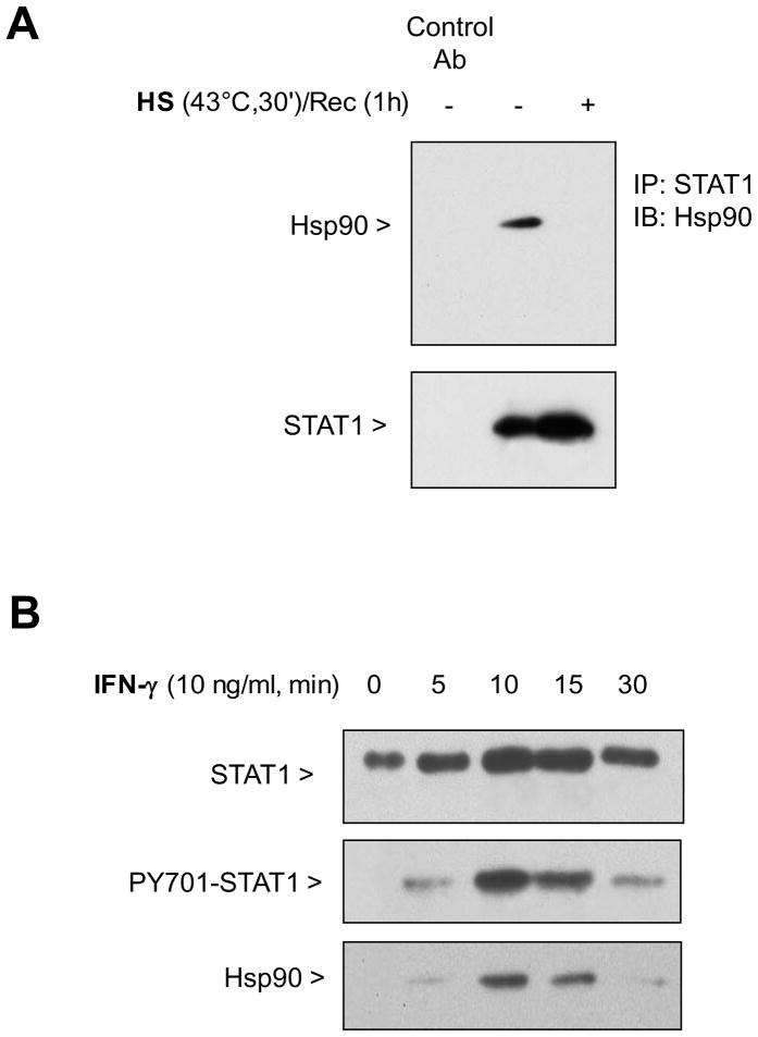
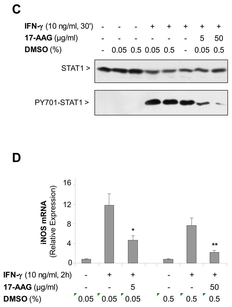
Figure 3. STAT1 forms a complex with Hsp90 enabling activation of STAT1 and is disrupted by SPR activation in alveolar macrophages.
Panel A. Heat stress causes a dissociation of the complex formed between STAT1 and Hsp90. MH-S cells were either untreated or were incubated at 43°C and allowed to recover for one hour. The cells were then lysed in a buffer containing 0.1% NP-40. Following centrifugation, the resultant supernatants were used for immunoprecipitation reactions using an antibody specific for STAT1. The resultant immunoprecipitates were examined via Western blotting for Hsp90 and STAT1. Panel B. Both STAT1 and Hsp90 are recruited to the plasma membrane upon stimulation with IFN-γ . MH-S cells were stimulated with IFN-γ (10 ng/ml) for the indicated times. Plasma membranes were isolated using dounce homogenization and differential centrifugation. The presence of total STAT1, phosphorylated STAT1 and Hsp90 was determined in the cell membrane fraction by Western blot. Panel C. Pretreatment with 17-AAG an Hsp90 inhibitor, inhibits phosphorylation of STAT1 in response to IFN-γ. MH-S cells were pretreated with 17-AAG an Hsp90 inhibitor , (5 or 50 μg/ml), one hour prior to IFN-γ stimulation (10 ng/ml for 30 min) and then analyzed for STAT1 and phospho-STAT1 by Western blot. Each set of experiments has been performed with the same final concentration of DMSO (solvent for 17-AAG). Panel D. Pretreatment with 17-AAG an Hsp90 inhibitor, inhibits IFN-γ-mediated iNOS mRNA expression. MH-S cells were pretreated with 17-AAG (5 and 50 μg/ml) one hour prior to IFN-γ stimulation (10 ng/ml for 2h) and analyzed for iNOS mRNA by real-time RT-PCR normalized with GAPDH mRNA levels. Each set of experiments has been performed with the same final concentration of DMSO (solvent for 17-AAG). Results are the mean ± SD of three experiments done in triplicate; *p < 0.05 from cells exposed to IFN-γ with 0.05% DMSO; **p < 0.05 from cells exposed to IFN-γ with 0.5% DMSO.


