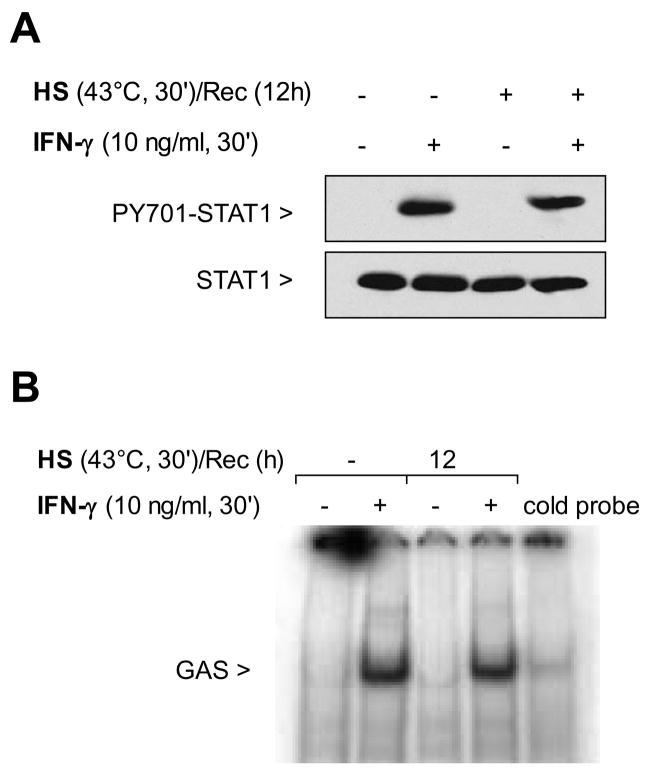
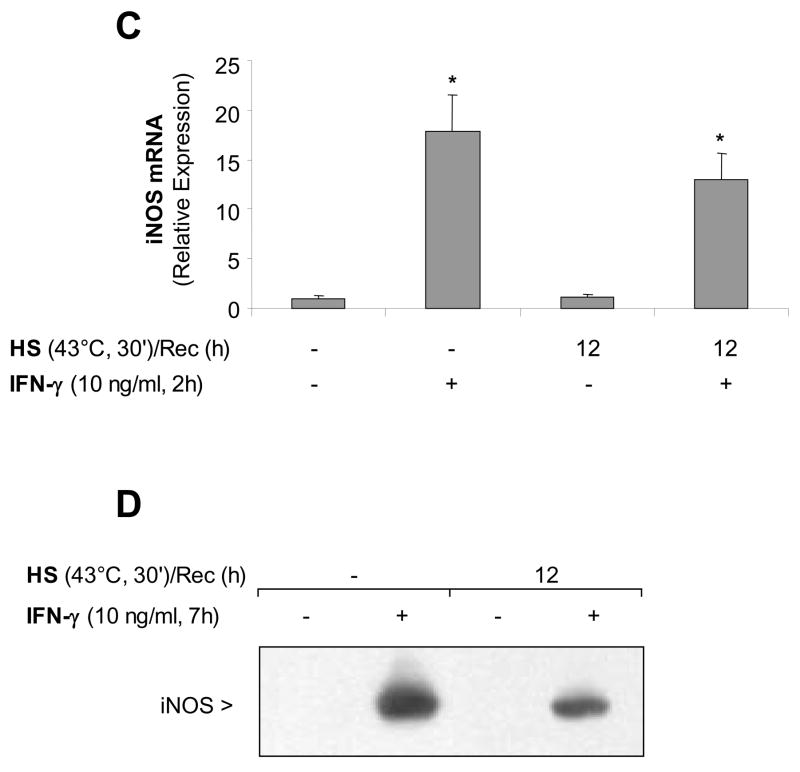
Figure 5. STAT1 signaling pathway is functional after 12 hours recovery post-heat stress induction and subsequent IFN-γ stimulation.
Panel A: MH-S cells were either untreated or were incubated at 43°C and allowed to recover for 12 hours prior to IFN-γ stimulation (10 ng/ml for 30 min) and then analyzed for STAT1 and phosphorylated STAT1 by western blot. Panel B: MH-S cells were either untreated or were incubated at 43°C and allowed to recover for 12 hours prior to IFN-γ stimulation (10 ng/ml for 30 min). Electrophoretic mobility shift assay (EMSA) analysis was used to examine STAT1 activity in IFN-γ-treated cells. Nuclear extracts were prepared (see methods) and 5 μg of nuclear protein were incubated with a 32P-labeled DNA containing STAT1 binding consensus sequence (GAS). In some reactions, a 100-fold excess of the unlabeled oligonucleotide was added (indicated as cold probe in the figure). The experiment was performed 5 times. One representative experiment is shown. Panel C: MH-S cells were either untreated or were incubated at 43°C and allowed to recover for 12 hours prior to IFN-γ stimulation (10 ng/ml for 2h) and analysis for iNOS mRNA by real-time RT-PCR normalized with GAPDH mRNA levels. Results are the mean ± SD of three experiments done in triplicate; *p < 0.05 from control cells. Panel D: MH-S cells were either untreated or were incubated at 43°C and allowed to recover for 1 hour prior to IFN-γ stimulation (10 ng/ml for 7h) and analysis for iNOS protein by Western blot; one representative experiment is shown, three additional experiments gave comparable results.


