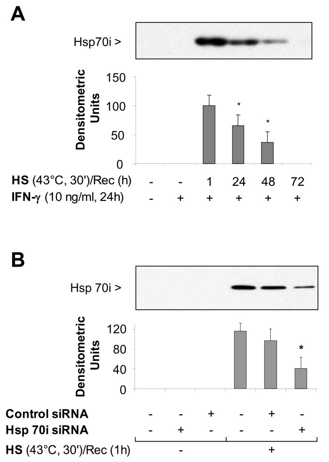
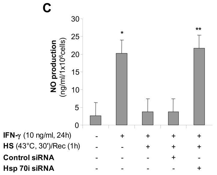
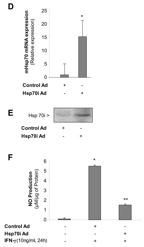
Figure 6. Inducible Hsp70 inhibits iNOS function due to activation of the stress protein response by heat.
Panel A. Time course of Hsp70i expression after heat stress. MH-S cells were incubated at 43°C for 30 min, then allowed to recover at 37°C for the indicated times prior to IFN-γ stimulation (10 ng/ml) for 24 hours. Cells were analyzed for Hsp70i by Western blot. Panel B. Transfection with a specific siRNA one hour after SPR activation inhibits the expression of the inducible Hsp70. MH-S cells were either transfected with a control siRNA or a specific siRNA to Hsp70 twenty four hours before heat stress (before) or heat stressed, allowed to recover at 37°C for 1 hour and then transfected with a control or a specific siRNA to Hsp70 (after). Twenty four hours post-heat stress, cells were lysed and the expression of Hsp70 protein determined by Western blot. Densitometry analysis results are the mean ± SD of three experiments; *p < 0.05 from cells treated with control siRNA. Panel C. Inhibition of heat stressed induced Hsp70 results in NO production in response to IFN-γ. MH-S cells were incubated at 43°C for 30 minutes allowed to recover at 37°C for 1 hour and then transfected with a control or a specific siRNA to Hsp70 (after). Twenty-four hours post-transfection, cells were exposed to IFN-γ (10 ng/ml) for 24 hours. Extracellular medium was assayed for nitrite using the Griess reagent. Results are the mean ± SD of 3 separate experiments with each condition carried out in triplicate; *p < 0.05 from cells exposed to IFN-γ alone; **p < 0.05 from heat stressed Control Ad infected cells exposed to IFN-γ. Panel D: Cells were infected with a recombinant adenovirus expressing Hsp70i. Forty-eight hours post-infection, cells were lysed and the expression of Hsp70i mRNA determined by real-time RT-PCR normalized with GAPDH mRNA levels. Results are the mean ± SD of three experiments done in triplicate; *p < 0.05 from control cells. Panel E: Cells were infected with a recombinant adenovirus expressing Hsp70i. Forty-eight hours post-infection, cells were lysed and the expression of Hsp70i protein determined by western. Panel F: Cells were infected as described above. Cells were exposed to IFN-γ (10 ng/ml) 24 hours post-infection for 24 hours. Forty-eight hours post-infection, the extracellular medium was assayed for nitrite using the Griess reagent. Results reported as NO production of infected cells. Results are the mean ± SD of 3 separate experiments with each condition carried out in triplicate; *p < 0.05 from control cells; **p < 0.05 from Control Ad infected cells exposed to IFN-γ.


