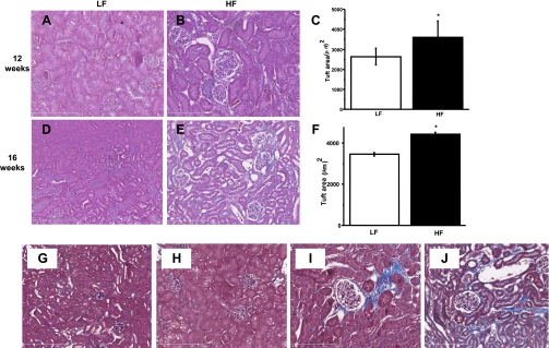
Fig. 2.
Glomerular hypertrophy and fibrosis, interstitial scarring, fibrosis, and mesangial space expansion in the HFD kidney. A–F: LFD-fed mice kidneys showed normal glomeruli structure at 12 and 16 wk (A and D), whereas kidneys in HFD-fed group showed gradually progressing increase in glomerular tuft size and Bowman's space expansion after 12 (B) and 16 wk (E). At least 30 glomeruli were measured in each group, data represent means ± SE; n = 30, *P < 0.05 vs. LFD. G: representative section of LFD-fed mouse kidney at ×100 with TriChrome staining, with normal glomeruli and mesangial cells at 12 wk. H: typical section of a 16-wk LFD-fed mouse kidney. I: representative of 12-wk HFD glomeruli with mesangial space expansion, fibrotic tissue, thickened membranes, and expanded Bowman's space stained in blue (×200). J: fibrosis, collagen, and interstitial scarring in a 16-wk HFD-fed mouse kidney (×200).

