Fig. 4.
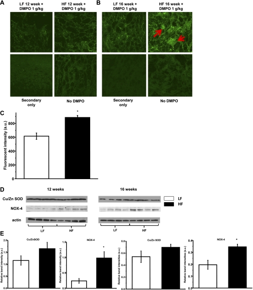
Cellular oxidative stress and protein radical formation progress over time on HFD feeding in the kidney. Animals were injected with the spin trap DMPO (1 g/kg, twice) before being euthanized, and then a DMPO-protein radical adduct-specific anti-DMPO antibody was applied for staining. A: minimal green staining can be observed in kidney sections after 12 wk of HFD feeding. B: protein radical formation was observed as positive green staining mainly in glomeruli after 16 wk of HFD feeding (red arrows). Kidney sections from non-DMPO-treated animals and omitting primary antibody on sections served as negative controls. C: evaluation of staining was measured as fluorescent intensity in glomeruli of the 16-wk group. Data show means ± SE; n = 15–20 glomeruli per each group. *P < 0.05 vs. LFD group. D: representative Western blots of Cu/ZnSOD and NOX-4 protein levels in LFD and HFD groups showing no change in Cu/ZnSOD levels, but a robust increase in NOX-4 levels on HFD. E: Western blot band intensities were evaluated in each group (n = 4). Data are expressed as means ± SE. *P < 0.05 vs. LFD group.

