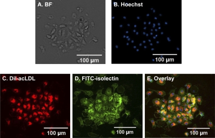
Fig. 1.
Phenotype characterizations of mouse bone marrow-derived endothelial progenitor cells (EPCs) after 7 days in culture. A: in bright field (BF), attached cells exhibit endothelial cell-like morphology and show their capacity to form endothelial colonies. B: Hoechst staining to detect the cell nucleus. C: cells show Dil-acLDL uptake. D: cells wee stained by FITC-isolectin. E: overlay of the 3 stains.

