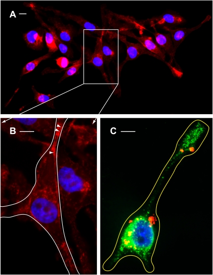
Fig. 8.
Morphological changes characterized by the formation of cell protrusions and redistribution of acidic vesicles as a result of aldolase knockdown. A: wide-field view of cells transfected with Alexa 555-conjugated siRNA-1 (red) followed by staining of actin cytoskeleton with Alexa 647-conjugated phalloidin (red) and nuclei with DAPI (blue). Long cellular protrusions (up to 20–40 μm) were clearly detected in all cells. B: enlarged image of box outlined in A. Arrowheads show cortical actin filaments. The border of a representative cell is outlined in white. C: epifluorescence microscopy merged image showing the aldolase knockdown phenotype characterized by cell shape remodeling accompanied by formation of long protrusions loaded with acidic vesicles. This representative cell was transfected with Alexa 555-conjugated siRNA-1 (red) followed by loading of acidic vesicles with DAMP and staining with Alexa 488-conjugated anti-DNP antibodies (green) and nuclei staining with DAPI (blue). The border of the cell is outlined in yellow. Scale bars all correspond to 5 μM.

