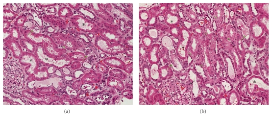
Figure 1.
Representative photographs of the renal biopsy showing tubular damage secondary to drug toxicity. In both panels (a) and (b) a number of tubules show moderate degree of acute tubular necrosis (asterisks). Some of tubules contain hyaline or epithelial casts in their lumina (green arrows), while several tubules show vacuolization of their cytoplasm (green arrowheads). One of the glomerular afferent arteriole shows swollen endothelia and occlusive change (black arrow).

