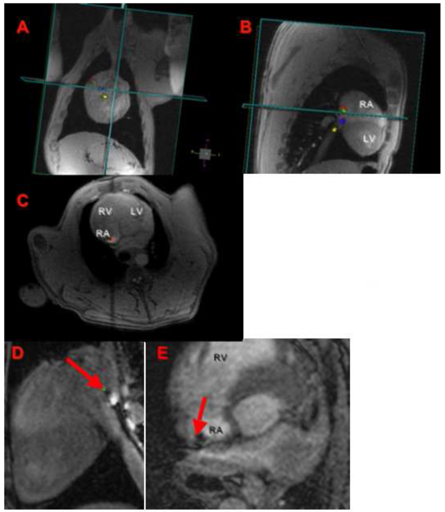
Figure 2. Assessment of catheter tip position and catheter tip-tissue interface by RTMRI and FLASH sequences.
An MRI compatible catheter is guided under RT-MRI (A–C) from the inferior vena cava to the lateral wall of the RA, this is seen in orthogonal planes (coronal, sagital, and axial views). The tip of the ablation catheter touching the atrial wall (red arrow) is seen in high-resolution T1w-FLASH images (D–E). RA: right atrium, LA: left atrium, RV: right ventricle, and LV: left ventricle.

