
Figure 1.
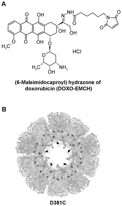
A. Chemical structure of DOXO-EMCH. B. Quaternary protein structure of the E2-D381C nanoparticle, comprising 60 subunits, viewed at the five-fold axis of symmetry. The Protein Data Bank crystallographic file (1b5s) is displayed using PyMOL (DeLano, 2002), and the doxorubicin conjugation sites (amino acid 381) are highlighted in black.