Fig. 3.
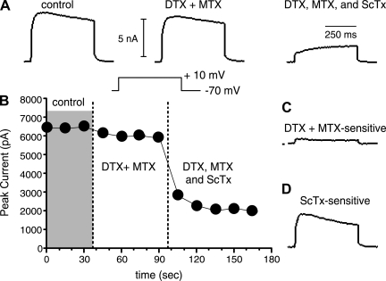
Pharmacological separation of slowly inactivating currents. A: representative traces (from a P23 animal) taken in control solution (left), after application of the Kv1 blockers 100 nM α-dendrotoxin (DTX) + 30 nM margatoxin (MTX) (center), and after subsequent application of the Kv2 blocker 600 nM Stromatoxin (ScTx) + DTX and MTX (right). Note that after the toxins a very slowly activating current remains. B: plot of peak current vs. time for the cell in A. Data were obtained from a holding potential of −70 mV. Voltage steps were to +10 mV. Currents were stable in control solution. DTX + MTX caused a small reduction in current. ScTx application blocked most of the remaining current. C: DTX+MTX-sensitive current (putative Kv1 mediated) obtained by subtraction of DTX+MTX trace in A from Control. In this cell there was a small transient and a larger persistent component to the putative Kv1-mediated current. D: ScTx-sensitive current (putative Kv2 mediated). This was the largest component in all cells.

