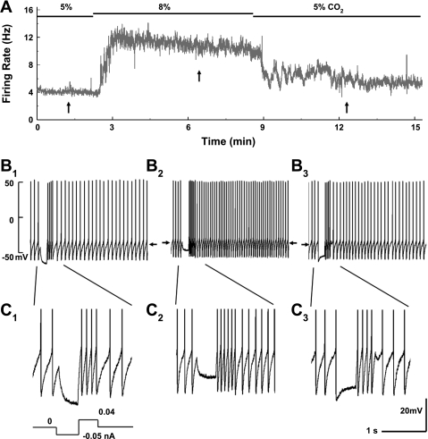
Fig. 1.
Whole cell recordings from locus coeruleus (LC) neurons in brain slice. A: neuronal firing activity was studied in instant-frequency histogram. The cell had a firing rate (FR) of 4 Hz at baseline with 5% CO2. Exposure to 8% CO2 raised the FR markedly. The peak of the FR (∼12 Hz) was reached in 1 min and then declined slightly throughout the 6-min 8% CO2 exposure. Washout with 5% CO2 led to almost complete recovery of the FR in 4–5 min. B: membrane potentials (Vm) and action potentials of the neuron. B1, B2, and B3 were obtained from A at the arrows from left to right, respectively. The Vm of each trace is indicated by the arrows. Hypercapnia augmented the FR and depolarized the cell (B2). The input resistance (Rm) was monitored by injecting a −0.05-nA current pulse every 20 s during the experiment and then calculated with Ohm's law, which is better seen in C with expanded displays. The Rm dropped by ∼50% during the high-CO2 exposure (C2). C1 and C3 are obtained from B1 and B2, respectively.

