Fig. 3.
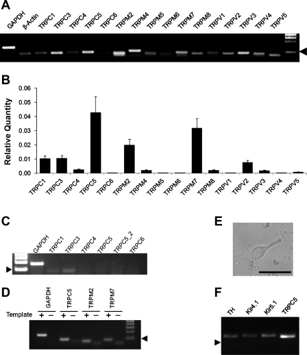
Expression of transient receptor potential (TRP) channels (TRPC) in LC neurons. A: all representative members in the TRPC, TRPM, and TRPV families were included in the RT-PCR analysis. Several TRP channels were clearly detected in the mRNA level. Arrowhead indicates 100 bp. B: analysis of TRP mRNA expression in the LC tissue with quantitative PCR indicated that the expression of TRPC5, TRPM2, and TRPM7 were severalfold higher than other TRP channels. Data are presented as means ± SE (n = 16–20 samples from 5 experiments). C and D: negative PCR controls. C: expression levels of various TRPC mRNAs in cardiac ventricular muscle. Of representative members in the TRPC families, only TRPC3 and TRPC1 mRNAs were detected in RT-PCR. Note that TRPC5 was studied with 2 different sets of primers, none of which produced a detectable band. Arrowhead indicates 100 bp (n = 3 experiments). D: another negative control for TRPC5, TRPM2, and TRPM7 expressions, the 3 most abundant TRPs in the LC (B). In the absence of template cDNAs, a weak and fuzzy band was found. The sizes of these bands were greater than primers, suggesting primer multimerization. In the presence of cDNA templates, a strong band was produced in each TRP, which was clearly larger than the primer band with expected size. More importantly, none of the primer bands was seen in the presence of cDNA templates. E and F: single-cell PCR analysis on the expression of TRPC5 in neurons. E: a cell acutely dissociated from the LC tissue. Calibration: 50 μm. F: single-cell PCR analysis showed that this cell was a tyrosine hydroxylase (TH)-positive neuron and expressed the TRPC5 mRNA. Inward rectifier K+ 4.1 (Kir4.1) and Kir5.1 mRNAs were also seen in the cell.

