Fig. 1.
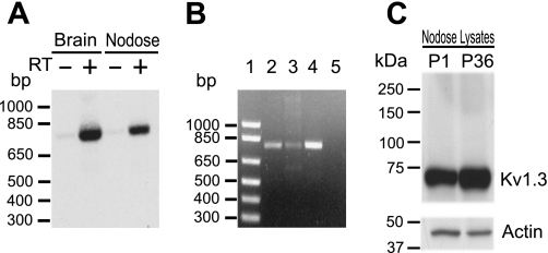
Detection of Kv1.3 α-subunits. A: PCR products resulting from the amplification of first-strand cDNA prepared with (+) or without (−) reverse transcriptase (RT) from the rat nodose ganglia (NG) or brain poly-A+ RNA with Kv1.3-specific oligonucleotides were separated by electrophoresis and transferred to nylon membranes. After Southern blot hybridization with 32P-labeled specific internal oligomers, the autoradiogram showed positive signals from the rat nodose and brain in +RT lanes and no signals in the control −RT lane. The oligonucleotide probes amplify cDNA of 792 bp. B: the specificity of the primers was tested using a sample of Kv1.3 cDNA. A single band of the predicted size (792 bp) was amplified from NG cDNA (lane 2), Kv1.3 cDNA (lane 3), and nucleus of the solitary tract (nTS) cDNA (lane 4). No PCR products were detected in the negative control lane (H2O; lane 5). Lane 1 is a 1-kb DNA ladder. C: Western blot analysis of Kv1.3 protein from the NG. The Kv1.3 receptor was identified in protein lysates from the NG dissected from postnatal day 1 (P1; 50 μg) and postnatal day 36 (P36; 20 μg) rats. A band of ∼65 kDa was detected in both lysates. Mobility of the protein correlated with the molecular size already reported for the Kv1.3 α-subunit (∼65 kDa). Western blot analysis also showed that Kv1.3 protein in neonates is less abundant than that in older animals.

