Fig. 2.
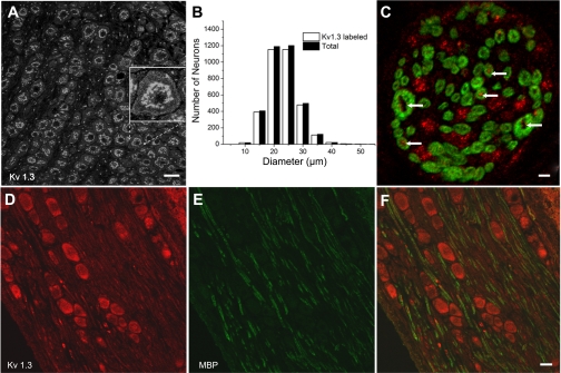
Immunohistochemical localization of Kv1.3 in the NG and aortic depressor nerve (ADN). A: anti-Kv1.3 (Neuromab) immunolabeling in postnatal day 30 Spraque-Dawley rat nodose slices. The image is a mass z-projection of five confocal sections acquired at 0.69-μm intervals. Calibration bar = 30 μm. The inset shows a zoomed image to illustrate weaker labeling near/at the membrane. B: histograms showing the broad distribution of anti-Kv1.3 labeling with the respective neuron diameter compared with the distribution of diameters in the total population. C: section of the ADN colabeled with anti-Kv1.3 (Alomone; red) and anti-myelin basic protein (MBP; green) to illustrate the presence of Kv1.3 in myelinated axons. Arrows indicate examples of the anti-Kv1.3 label. These results were obtained in the ADN from two animals. Note the presence of only two large-diameter axons. Similar results (2–3 large axons with diameters > 2 μm) were obtained in five other ADNs. Scale bar = 2 μm. D and E: images of 8-μm nodose sections colabeled with anti-Kv1.3 (red; D) and anti-MBP (green; E). The overlay (F) shows the presence of Kv1.3 antibody in myelinated and unmyelinated axons in the ganglion. Scale bar = 20 μm.

